Monday, 6 April 2015

How Does Penicillin Work?

ANTIBIOTICS

The first antibiotic, penicillin, was discovered serendipitously in 1928 by Alexander Fleming. A medic by training Fleming went on to specialise in bacteriology before leaving his comfortable lecturing position for the trenches of World War I. He served as a medic in French field hospitals on the western front. There's no doubt that during his military service Fleming was exposed to the horrific toll that bacterial infection caused wounded troops. Many amputees died from non-life threatening surgery due to infections obtained in unsanitary field hospitals. But the discovery of penicillin revolutionised surgery, allowing patients to recover from infections that could otherwise kill them.
Alexander Fleming, perhaps not the happiest scientist there ever was. 

So effective was this wonder drug that Fleming was awarded the Nobel prize for Medicine in 1945. By this point, penicillin had saved thousands of lives both on and off the battlefield.

So how does Penicillin work? Well, to understand that one needs to understand the basics of cell biology. Cell biology studies with what cells are made of, and how they operate. A detailed understanding of cell biology is beyond the scope of this post, but it is useful to understand bacteria in the context of other forms of life. 

TYPES OF LIFE

It might seem surprising, but every living thing on the planet can be categorised into just there different branches on the complex tree life. These branches of life are called Prokaryotes, Eukaryotes, and Archaea. In addition there are also viruses. Viruses are often difficult to introduce when discussing simple life-forms because while they are much more simple than a bacteria cell, they are not always considered living cells. They have many of the characteristics of life, movement, sensitivity to their surroundings and so on, but unlike living cells they are unable to reproduce without a host cell. That is, they rely on infecting a bacteria or human cell to make more copies of themselves. For this reason, viruses are often considered nothing more than assembles of biomolecules, albeit, very interesting and often dangerous assemblies of biomolecules.
THE ARCHAEA

The Archaea branch of life contains the simpler organisms, the early forms of bacterial life on earth. They are single celled with an amazing ability to survive and adapt, often living in environments that we would consider hostile. For example, there are some Archaea bacteria that live in the very darkest depths of the ocean, in close proximity to hydrothermal vents. These living conditions contain extreme pressures and temperatures and there no sunlight. However, this proves to be no obstacle to these hardy organisms who thrive on the constant supply of noxious chemicals released from deep below the surface of the earth. 

THE PROKARYOTES

Like the Archaea, these are simple, unicellular organisms, but they tend to live in less hostile environments. These are the bacteria you are more likely to have heard of. The E.coli bacteria associated with food poisoning for example, or MRSA infections commonly contracted in hospitals. Prokaryotes are tough, adaptable, and capable of reproducing quickly making them potentially very dangerous. However not all of them cause disease in human (a quality known as pathogenicity). Some are actually beneficial to us, bacteria in our intestine actually help us digest food we eat, and produce vitamins for us in return.

THE EUKARYOTES

Finally, the eukaryotes. These are the most complicated organisms, and include some unicellular life such as yeast cells, but also the more complicated multicellular organisms. This includes everything from sponges to worms to humans. The individual cells that make up a human are much more complicated than an individual bacterial cell. This complexity comes at a price, namely the speed at which a human cell can make more of itself. Bacteria are very quick to reproduce but human cells, by comparison, are slow but make a more faithful copy of themselves. 

THE ANATOMY OF BACTERIAL CELLS

Bacteria themselves are incredibly interesting lifeforms. Although more simple than us they are  still very complex. They consist of a bunch of cell organelles, and some DNA wrapped up in a fatty skin called the cell membrane. Surrounding this cell membrane is a tougher shell called the cell wall. The cell wall is made of a tough material called peptidoglycan, and offers the bacteria some protection from the outside world. You can think if it as multiple layers of wire mesh; rigid, strong, but also flexible.

The anatomy of a bacterial cell. The yellow "skin" on the outside represents the cell membrane, and the cell wall. 

This peptidoglycan mesh is a composite of sugars and amino acids. The sugar components have the unwieldy name of N-acetylglucosamine, and N-acetylmuramic acid so for ease, biologists abbreviate these to simply NAG and NAM. NAG and NAM sugars are joined together in a chain, and individual chains become cross-linked with small protein molecules for extra strength and rigidity. As with many processes in biology the formation of a bacterial cell wall is facilitated by an enzyme, in this case, an enzyme called transpeptidase.
This image shows the function of the transpeptidase enzyme in making bacterial cell walls. The red and orange hexagons represent the individual NAG and NAM sugars that combine in long chains. The chains have small pieces of sticky protein (peptides)  hanging from every NAM molecule. The transpeptidase stitches each NAG-NAM chain together by fusing the peptides molecules together. These fused chains are the basic structure of a bacterial cell wall. 

Bacteria are constantly making and degrading their peptidoglycan cell walls during the process of cell division. It's a necessary part of how the divide and make more of themselves. Without the ability to make and repair their cell walls bacteria become flooded with water from outside the cell, and burst. A process called cytolysis. Therefore, the transpeptidase enzyme is essential for the survival of bacterial cells. Some antibiotics work by inhibiting the action of this essential enzyme. But how?

THE STRUCTURE AND FUNCTION OF ANTIBIOTICS

There are many different types of antibiotics, and they can be classified on the basis of their chemical structure, mode of action, or spectrum of action. As mentioned, Penicillin works by inhibiting bacterial cell wall synthesis, but other antibiotics work in different ways. What is it about penicillin that makes it work the way it does? Well, like a lot of biochemistry, it all comes down to to the atomic structure of the penicillin molecule. Fleming did not know this at the time of his discovery, but penicillin actually looks very like the peptide molecules used to strap the peptidoglycan chains together.


So, when we take penicillin to treat a bacterial infection we are presenting the bacterial transpeptidase enzyme with a choice. The enzyme can either, grab hold of the peptide molecule (D-Ala-D-Ala) like it's supposed to, or it can grab a molecule of penicillin. If it grabs a molecule of D-Ala-D-Ala then everything goes to plan, at least as far as the bacteria are concerned. Cell wall biosynthesis occurs normally, and the bacteria happily go about reproducing. However, if the transpeptidase enzyme grabs a molecule of penicillin instead of D-Ala-D-Ala, then the bacteria are in trouble. Penicillin will bind strongly to transpeptidase enzyme, effectively gumming up the works. We now have a situation much more like the image shown below.

Here, the penicillin molecule is represented as the yellow and black circle, jammed in the mechanism of the transpeptidase enzyme.

Here, the penicillin molecule is represented as the yellow and black circle, jammed in the mechanism of the transpeptidase enzyme. With penicillin gumming up the works it is not possible for transpeptidase to do it's job. The more penicillin we take the more this situation occurs, making it very difficult for the bacteria to reproduce. This mechanism of actions applies only to penicillin and antibiotics that look like penicillin such as amoxicillin. Different antibiotics work in slightly different ways, targeting different proteins that are important for bacterial cell survival.

ANTIBIOTIC RESISTANCE

Antibiotic resistance is becoming an increasing problem. It refers to the ability of some bacteria to keep reproducing despite the presence of antibiotics designed to kill them. So, how have these crafty bacteria overcome our sophisticated chemical weapons? The answer is evolution. Every generation of bacteria results in a slightly new organism. It's still a bacteria, but it's ever so slightly different from the "parent" that made it. Sometimes these differences are enough to result in interesting properties such as antibiotic resistance.

Antibiotic resistance stems from an overuse of antibiotics in medicine. This could be over-prescription by doctors, or people administering for diseases not associated with bacteria, such as colds, or flu, caused by viruses.  For any given infected individual it is unlikely that a single dose of antibiotics will wipe out every single bacterial cell in the body. Any cells that are left will produce offspring which become resistant to their hostile environment, and thrive in their new competition free zone. Next time these bacteria encounter this antibiotic they have already adapted to it, and produce their own chemical weapons to destroy ours.

CONCLUSION

So, there you have it. Penicillin works by preventing the construction of bacterial cell walls, causing bacteria to explode via an intake of water. Penicillin is just one of many types of antibiotic, which can produce their effects in different ways. Bacteria are adapting to these chemical weapons by producing chemical weapons of their own, destroying of the antibiotics before they can take effect. This has resulted in an literal arms race between us and the bacteria, but we are losing. The result is multidrug resistant bacteria such as MRSA which is having a pronounced effect on hospital patients worldwide. If we are to win this arms race we need think differently, and stop relying on brute force approaches. 


Friday, 19 December 2014

Genetically Modified Organisms

BACKGROUND
This is a post about genetic modification, and genetically modified organisms, or GMO's for short. Using farming as an example I will discuss how genetics can be used to improve traits such as milk production in cows, and disease resistance in potato crops. The aim of this post is to show how genetics has some real-world applications that affect all of us.

THE ORIGINS OF GENETICS
Although farmers across Europe have been breeding their cattle and crops selectively for centuries, it was a nineteenth century Augustinian monk who first realised the power of genetics. Friar Gregor Mendel used his monastery's pea garden to study the size, shape and colour of pea plants and their seeds. This unassuming research allowed him to identify the root of how individual traits are passed from parents to offspring, a term now named in his honour as Mendelian inheritance. The term “Mendelian Inheritance” refers to the transfer of genetic material, DNA, from parent to offspring. Centuries of research since Mendel’s humble beginnings in his pea garden has shown that many characteristics of plants and animals are determined by small packages of DNA called genes.  I have discussed, genes, and DNA in detail in a previous post so I won't go into detail here.

THE IMPORTANCE OF GENETICS
In the 150 years since Mendel our understanding of genetics has developed enormously, and modern scientists have now gained more precise control over the characteristics that can be added or removed from a variety of plants and animals. With the help of Teagasc, an independent state-funded agricultural research body, we are moving away from older and more costly methods of crossbreeding. Modern genetic manipulation now means that desirable traits can be selected for while undesirable ones can be left behind. This means that today’s farmers can save both time and money crossbreeding cattle and crops and increase crop yields for an ever expanding food market.

WHAT ARE GMOs?
Genetically modified organisms, or GMOs are organisms which have been altered through the addition or removal of individual genes. Human beings have been living in harmony with genetically modified food since the beginning of agriculture in the form of disease-resistant crops or bulkier cattle for better meat production. Although sensationalised media reports sometimes paint GM food as unnatural “Frankenstein food” that may pose a risk to our health or the environment, the reality is much less sinister. In fact, modern genetic modification techniques allow farmers to better control desirable gene inheritance in a fraction of the time traditional methods would have required.

CASE STUDY 1: THE HOLSTEIN-FRIESIAN/NEW ZEALAND JERSEY MIX
In 2013, for the first time in our history, Irish food and drink exports approached €10 billion and the dairy industry accounted for almost a third of this. Dairy exports in 2013 amounted to €3 billion, up 15% from 2012, and are expected to rise again this year. This is a testament to the high standards Ireland sets and maintains in this sector. The majority of Irelands milk production and calving is seasonal, with over 75% of milk production occurring from April to September, whilst 79% of calves are born between January and April.

The Irish dairy herd is dominated by the Holstein-Friesian breed, which makes up 63% of the national herd. Our farmers favour this breed because of its exceptional milk yields, a characteristic arising from breeding programmes in the US. When we consider that Ireland has a limited calving season of only four months, we can see that improving our herds reproductive success would have a knock-on gain in the milking season and allow farmers to boost their profit margins. Until recently, any potential gains to be had from cross-breeding the Holstein-Friesian with a more fertile breed were disregarded by farmers wary of tampering with the Holstein-Friesian’s milk yield. But all that changed when the Holstein-Friesian was crossbred with the New Zealand Jersey.
The Holstein-Friesian (left)  and New Zealand Jersey (Right)
Although the New Zealand Jersey has slightly inferior milk production properties compared to the Holstein-Friesian, it does have superior reproductive qualities and tends to live longer. A recent Teagasc study showed that the offspring of the Holstein-Friesian and New Zealand Jersey, called F1, had the characteristics required to improve the overall profitability of the Irish herd.
The results of the Teagasc study, with all values displayed relative to the Holstein-Friesian breed.
The Teagasc study revealed that the quantity of milk produced by the F1 crossbreed was marginally lower than the Holstein-Friesian, but this issue was compensated for with higher fat and protein content. The results also showed a modest improvement in turnaround time from calving to conception compared to the Holstein-Friesian. On top of this, the significantly hardier body condition of the F1 means lower maintenance costs for the farmer. When we add up the findings of this study, it clear that the benefits far outweigh the negligible disadvantages of crossbreeding these two strains. It’s no wonder then that New Zealand has already begun adopting this breed, with the F1 making up 33% of their national herd.

CASE STUDY II - GENETICALLY MODIFIED POTATOES
Potatoes are a staple ingredient on dinner tables all over Ireland, but it’s hard to mention this delicious starch without thinking about the Great Famine of 1845. This tragic episode in Irish history was caused by a fungus called Phytophthora infestans, commonly known as potato blight. Incredibly, over 150 years on, blight continues to be a serious concern for present day potato farmers.

In the battle against blight, fungicide remains our main weapon of choice. Fungicide is effective, but it requires as many as 15 to 20 sprays a year to secure a good potato crop. European regulations on fungicides are becoming more restrictive and, although many welcome this decision from an environmental perspective, potato farmers will need new weapons to protect their crop. As yet there are no blight-resistant strains of potato in Ireland but genetic modification may hold the key.

Some Central America potato strains possess genes known to provide protection against blight. Traditional methods of cross-breeding potatoes are both time consuming and inefficient, but new genetic modification methods have had great success in introducing these genes to European strains of potato. Teagasc collaborations on an international level have led to possibility of blight-resistant potatoes being developed in Ireland. The picture below highlight the significance of their efforts.
The image on the left shows a GM potato line (A15-031) containing genes for blight-resistance, while that on the right shows a unmodified potato crop
The effects of blight on both strains can be seen, with the GM potato crop remaining healthy despite the presence of blight, while the unmodified crop succumbs to the disease. This development provides farmers with new options going forward. However Ireland proceeds, the decisions made by this generation is likely to have very real and long-lasting consequences to future generations.

CURRENT OPINION GMOs
As with any new technology, a decision needs to be made on how and when it should be implemented. Those in favour of GMOs will urge for an early adoption to get ahead of European fungicide restrictions as well as to take advantage of any financial gains that can be made. Those who favour caution will prefer to wait and see how GMOs pan out for other nations. Teagasc is very clear about its stance on the issue of GMOs, making sure to distance itself from the commercial profit-oriented organisations and instead favouring unbiased publically-funded research. Dr. Ewen Mullins, Senior Research Officer of the Crop Science Department at Teagasc says, “We can’t rely on research done outside of the country by groups that are either for or against the technology”. Ireland has a lot to gain from probative research like this, but we need to be sure we are acting on the best available evidence. Teagasc is cautiously optimistic about introducing this GM potato crop in the field, stating, “Arising from the preliminary study completed in 2012, we now know that the GM potato variety we are researching has the potential to resist Irish blight strains but much more work is required and this will commence in 2013.”

THE FUTURE OF GMO IN IRELAND
Genetic modification will play an ever-increasing role in the growth and development of Irish agriculture. Ireland is fortunate to have a unique blend of traditional farming experience and a highly-developed scientific sector, allowing us to integrate old and new expertise to our benefit. The ability to skillfully handle GMOs will mean we can find tailor-made solutions to the various challenges facing the Irish agricultural sector. We can only speculate what Mendel would make of these modern advance, but I’d think he’d agree it’s not exactly “easy peasy” work.

REFERENCES


Thursday, 20 November 2014

Caffeine - What Is It, And How Does It Work?

BACKGROUND
This blog post is designed to give you basic information about caffeine. Why? Well, caffeine is an interesting compound. It is a legal drug, socially acceptable and cheap to buy. It is also frquently in the media, being linked to diseases such as Cancer and MS, but it is also associated with cognitive enhancement of memory and concentration. It's always worthwhile to question whether there is any validity to such claims: could caffeine possibly have any influence on concentration or memory for example? I will attempt to address some of these questions in future posts, but for now this is simply an introduction to what caffeine is, what it does and how it does it.

CAFFEINE
Caffeine is a small chemical compound with stimulant properties. Stimulants include a range of interesting compounds such as the amphetamine compounds MDA, MDMA, and methamphetamine. All stimulants promote increased levels of alertness, increased reaction time, and some degree of euphoria. Unlike amphetamine based stimulants however caffeine does not have psychedelic effects. Caffeine occurs naturally in the seeds of coffee plants, tea plants and in the cocoa beans, used to make chocolate. It is also added to synthetic products such as energy drinks, or painkillers where it is used to amplify their pain-relieving effects.

CAFFEINE IN THE BODY
Once ingested caffeine rapidly enters the blood stream. From here it makes it's way to the brain, and stimulates the cells of the central nervous system, (CNS). Approximately 12 hours after ingestion the caffeine molecules have been eliminated with the help of liver enzymes. In fact, the elimination of caffeine from the body is very complicated and involves a single molecule of caffeine undergoing 24 distinct modifications to its structure before it can finally excreted in the urine.

CAFFEINE AND ADENOSINE
Chances are you have never heard of adenosine, but to make sense of how caffeine interacts with the body I need to introduce and discuss the role of this new molecule. Adenosine is a small chemical compound called a purine nucleoside. It occurs naturally in our bodies and is involved in a number of important functions.
The exact function of adenosine  depends on where it is the body. For example, adenosine in the heart is required for maintaining a regular heartbeat, but it's also required for blood vessel dilation, smooth muscle contraction, neurotransmitter release, and metabolism of fat. In fact, the effects of adenosine on the heart are exploited in medicine where it is used to treat instances of supraventricular tachycardia. Interestingly, adenosine is also responsible for regulating sleep. I will go into more detail on this below.

ADENOSINE AND ATP
Our brain utilises an enormous amount of energy in the form of a chemical called adenosine triphosphate, or ATP. In the same way that ashes are the waste product of burning coal, adenosine is the waste product of burning ATP. Subsequently, as we continue to utilise our brains throughout the day, the levels of adenosine continue to rise. Rising levels of adenosine in the brain result in a person feeling tired, and ultimately going to sleep. While sleep still uses a lot of neural activity in the form of dreams, the rest of the body is on standby, giving it a chance to replenish its stocks of ATP.
CAFFEINE AND THE ADENOSINE RECEPTOR
Any molecule that interacts with a receptor in the brain is said to have a psychoactive effect, and this includes naturally occurring compounds such as dopamine and serotonin, associated with mood, movement and appetite, or acetylcholine associated with memory formation. Both adenosine and caffeine molecules mediate their psychoactive effects by binding with a specific protein molecule called the adenosine receptor, or ADR for short. Receptors are protein molecules found on the outside of cells where they act mediators between the inside of the cell and the outside environment. Receptor proteins are the primary way in which cells obtain information from their surroundings.

THE ADENOSINE RECEPTOR (ADR)
The ADR receptor is found in different regions of the body, primarily the brain, but also in the heart. The structure of this receptor molecule with caffeine bound to it is shown below. The images below show the ADR, from the front and top. The last image shows a zoomed in shot, with some of the protein cut away to show caffeine molecule more clearly. You'll notice the receptor protein is quite large, and somewhat tubular in shape. This tubular shape allows it to position itself in the cell membrane, an ideal location for relaying messages between the inside and outside of the cell. Notice, the caffeine molecule, shown in yellow, binds at the top of the receptor protein. All of the effects of caffeine, alertness, changes in heart-rate etc, are the result of caffeine binding this receptor protein. The binding of caffeine to this receptor instigates a whole new cascade of events that propagate the effects of caffeine all the way through the body and brain. The details of this cascade are too tricky to explain here, but for the moment all you need to know is that the result of adenosine binding to the ADR is that neural cell activity is slowed in preparation for sleep. Caffeine interferes with this system by binding to the same location as adenosine does, essentially blocking access to the ADR, and preventing adenosine from doing its job.
NEUROANATOMY 101
Lets imagine the brain is a vast computer network of CPUs and ethernet cables. In the brain, the ethernet cables are called neurons, and the CPUs are the nucleus of those neurons. There are some similarities between these two systems, they both have a bandwidth associated with them, i.e, a maximum rate of information transfer. They both have insulation to prevent loss of the signal, as well as eliminate cross-talk between wires, and in both systems information is rapidly transferred in discrete packets. For Ethernet cables this is a purely electrical signal, but for neurons this is mix of both electrical and chemical signalling.
Image on the left shows an artists representation of neurons, brain cells. The long tendril like structures are called dendrites. The synapses are the gaps between connections, where the electrical signal becomes a chemical signal. This is analogous to ethernet cables, right, which also carry electrical information, are insulated, and connect to each other via switches/synapses.
CHEMICAL SIGNALLING
The long tendril like structures of neurons are called dendrites. Dendrites are responsible for information transfer in the form of small charged molecules moving rapidly through the cells. The interface between two dendrites is called a synapse. The synapse is the region where electrical signals become chemical signals. If you want to interfere with information transfer in the brain the synapse is one of the places you can do it. This is where many of the commonly used drugs mediate their effects.

STIMULANTS AND INHIBITORS
To regulate all the traffic that occurs between neurons, the brain releases a mix of stimulant and inhibitor molecules. Both types of molecule occur naturally, being made by the brain cells themselves. Stimulants promote signalling between neurons, while inhibitors slow it down. Together stimulants and inhibitors provide a feedback mechanism, so when energy stores in the body are low we attenuate our neural activity accordingly. If you drink a cup of coffee, the caffeine molecule takes the place of adenosine at the ADR, and the signal that normally comes from adenosine that says "slow neural activity" is now missing. This results in a brain without "brakes" as it where and we continue to fire off signals from our neurons despite the need for  rest. By ingesting caffeine we have essentially hijacked our own brains and decided to over-ride our own internal feedback mechanisms for determining when we should rest. This is pretty cool in my opinion.

WAIT..I'M CONFUSED
Listen, I don't blame you. In order to discuss this seemingly simple molecule, caffeine, I had to introduce a lot of concepts and terminology. Biology is complicated like that, there are lots of interactions occurring all the time.  It is a complicated topic, but here is a summary that should help.
  • Caffeine acts a stimulant by preventing the action of adenosine at the adenosine receptor protein. 
  • This results in a removal of the "brakes" in the brain, and all the stimulatory neurotransmitters in the brain are free to roam around, and stimulate.
  • This translates into the physical characteristics we associate with coffee/caffeine, such as increased heart rate, increased reaction time, and increased concentration. 
As for any of the detrimental side effects associated with this, well, that's more difficult to determine. But there is plenty of media coverage on the topic. The links between caffeine and diseases will be explored in another blog post. In the meantime you can see for yourselves how common a news story this is, as the links below demonstrate, even a short time on the BBC news website shows how much we enjoy hearing about this topic.

ADDITIONAL READING


Wednesday, 8 October 2014

Proteins: Why They're Cool

BACKGROUND
The purpose of this post is not to provide specific information of a vast array of proteins but rather to give you an overview of what a protein is, and why they are important. It is to act as a reference point for later posts which will refer to terms like amino acid, enzyme, and protein structure, and to give an appreciation for the complexity of these molecules. As cool and interesting as the study of DNA, genes and genetic disorders is, the study of protein biochemistry is much more fascinating. Just as there are diseases associated with mutations in DNA, there are diseases that result from the poor organization of protein molecules. You will have heard them in the form of BSE, CJD, and Alzheimer's. I plan to discuss these in more detail in later posts, but for now, here is an introduction to proteins, and why you should care what they are.

PROTEINS
Proteins, along with lipids, sugars, and nucleotides, are one of four macromolecules of life responsible for many vital functions within a cell. There are three major classifications of proteins, structural, enzymatic and cell signalling/receptor proteins. These are distinguished by both their structure and their function, and both structure and function are intertwined. That is, the shape and composition of a protein really matters, and determines how it behaves in the cell. 

COMPOSITION
Proteins are composed of small chemical compounds called amino acids. There are a total of twenty amino acids, all similar in size and composition, but each of them unique. Amino acids combine to form complex and unique three-dimensional protein structures. When two amino acids combine they form what is called a dipeptide. When many amino acids combine they are called a polypeptide. Proteins are composed of polypeptide chains which fold into three dimensional shapes. The image below shows the chemical structure of each of the twenty amino acids. The point of this is not to confuse you with chemical structures, but instead to get an appreciation for the complexity and variety of these compounds. If you look at each of these compounds you will notice they all share the same chemical structure, except for the parts highlighted in red, these are called side chains.
Image taken from http://amit1b.wordpress.com/the-molecules-of-life/about/amino-acids/
The amino acid side chains are partially responsible for providing a protein with its shape, and function. Like DNA, each compound is given a one letter code so that it can be referred to quickly and easily, and proteins can be depicted as long sequences of letters. For example, the amino acid Isoleucine is "I", Tyrosine is "T", Alanine is "A", Serine is "S", Valine is "V", Asparagine is "N" Cysteine is "C", Lysine is "V" and Aspartate is "D". A peptide composed of these amino acids would be depicted as ITASVNCAKKIVSD. You'll notice the same amino acid can be present more than once in the same polypeptide sequence.

STRUCTURE
Biologists love to identify and classify things, and this extends all the way down to the molecular level. This linear arrangement of amino acids depicted in the previous paragraph is called the primary protein structure. The primary structure folds in on top of itself to form one of two new structures called (i) the alpha-helix, or (ii) the beta-sheet. Sheets and helices are often linked together by smaller segments of the protein which do not assume any three dimensional shape.
Three dimensional structure of a helix (left) and sheet (right). Image was made from the data on a small protein called hen egg white lysozyme, using the rendering program YASARA.
Within a single protein molecule there could be numerous helix and sheet structures linked together by small unstructured regions. These helix and sheet structures are called secondary structures, and the primary amino acid sequence determines which one is formed since certain amino acids have a propensity for helix formation, while others favour sheet formation. The combination of secondary structures linked together by small unstructured regions of protein is called tertiary structure, and is generally what is being referred to when biologists talk about proteins. The process of going from primary to tertiary structure is referred to as protein folding, and it's a fascinating aspect of protein biochemistry. See here for an excellent video of how proteins assemble from their component parts.

FUNCTIONS
Like many other aspects of biology, there is a structure/function relationship with proteins, by which I mean the function of a protein is dependent on the structure and composition of the protein. Proteins are roughly separated into enzymes and structural proteins. They both have vital but distinct roles in the body. Enzymes modify and change things in the body, whereas structural proteins provide physical support to the shape of a cell.

PROTEINS IN THE CELL MEMBRANE
Proteins are present within every part of the cell in addition to the cell surface. Proteins on the cell surface are typically involved in cellular communication, as well as transport of materials in and out of cells. Below is the structure of a cholesterol transporter (shown in yellow), imbedded in the cell surface, a region referred to as the lipid bilayer, or cell membrane. Even without knowing anything about the structure of cells in general you can see how this protein carves out a space in the lipid bilayer and provides a channel that allows the passage of small chemical compounds. While some compounds would be able to make their way through the lipid bilayer without a protein channel others would never stand a chance.
Image taken from Ukasz Jaremko, M. Jaremko, K. Giller, S. Becker, M. Zweckstetter. Structure of the Mitochondrial Translocator Protein in Complex with a Diagnostic Ligand.Science, 2014; 343 (6177): 1363 DOI: 10.1126/science.1248725
PROTEINS INSIDE CELLS
Proteins inside cells perform a massive array of functions. For example, the conversion of food to cellular energy involves ten proteins (enzymes), in addition to a transporter protein located on the surface of the cell. Each one of these enzymes performs a small but necessary part in the conversion of sugar to ATP, and this is just one small metabolic pathway in the body. There are proteins for carrying oxygen around the blood, proteins for regulating the formation and release of dopamine, and serotonin in the brain, proteins for clotting your blood and proteins for regulating how and when your DNA is used.

Actin is one of the most abundant proteins in our cells, and forms what is referred to as the cytoskeleton. In the same way that we have skeletons made of bone that allow us to move, cells have cytoskeletons make of long filaments of actin protein that allow them to move. The structure of actin is shown below. As you can see, The actin protein is long, and thin, exactly what you need for providing structural support to a cell. Multiple actin molecules bundle together in close proximity to provide additional tensile strength.
Actin filament, image was made from X-ray crystal structure data found in the protein data bank, PDB ID 1M8Q.
Compare this to the structure of some other non-structural proteins shown in images (a) to (d) below. These proteins are enzymes, or transporters, and they do not need to be mechanically tough. They move around the cell, or bind to substrates which often induce a change in structure. You can immediately see the difference between these proteins and the actin filament protein.
The structure of four proteins not involved in structural stability of the cell, (a) hen egg white lysozyme, an anti-baterial protein, (b) hexokinase, a protein involved in glucose metabolism, (c) calmodulin, a protein involved in calcium transport, and (d) haemoglobin, a protein involved in oxygen transport. All images made using YASARA.
SO - THERE ARE DIFFERENT PROTEINS, SO WHAT?
Well, my point here is that when people come across the word protein they tend to think of something pretty uninteresting, a block of cheese, or some milk, or maybe some beige coloured protein powder. It's true, these are foodstuffs that contain proteins, but what I have a attempted to show you is that proteins are far from boring to look at, and far from uninteresting. They are complex three dimensional structures made of smaller simpler components. The can assume a vast array of shapes, and the shape and function are linked.

It's worth noting, cells exist as a collective, they need to share information about their surroundings. Cells need to know when to divide, when to stop dividing, when to signal for an immune response, and when to metabolise sugar to energy. All of these functions are performed by protein molecules on the surface of the cell, or embedded in the cell membrane. You can think of them as a mixture of radio transmitters/receivers controlling a set of locked gates. Nothing gets past unless it has the correct chemistry to do so.

If you think of DNA as the instruction booklet for the manufacture of a human being, proteins are the machines that create and destroy all the components, they ferry raw materials around, they make sure there is a constant supply of raw material around, they regulate production to meet demand, and they work together to fight infection, heal wounds, and keep us breathing. They really are the cogs in the machine that is our body. 

REFERENCES/EXTRA STUFF
  • YASARA, short for Yet Another Scientific Artificial Reality Application, this is a free application for downloading and viewing protein molecules called PDB structures.
  • Check out this cool video from the PDB on protein structure 
  • The protein data bank, a website for downloading PDB files, crystal structures, of proteins for viewing on your own computer.

Wednesday, 3 September 2014

Hemochromotosis - Part 1 - Introduction & Genetics

BACKGROUND
I have a disease called hereditary hemochromotosis, abbreviated HH for short. However, I know next to nothing about it despite being diagnosed nearly a year ago. This is my fault, since I've left it up to the public health service in Ireland to look after me, and really I'm not an urgent case. However, just because I am not dying doesn't mean my life is not adversely affected. In actual fact, I don't know how much my life is being adversely affected, so I am going to use my blog to teach myself and others about this disease. It's also a generally interesting insight into the genetics of an interesting metabolic disease.

WHAT IS HEMOCHROMOTOSIS?
Hemochromotosis is a disease that results in abnormally high levels of iron in the blood. It is a genetic disorder, meaning it is not acquired as a result of infection from bacteria or viruses for example, and is not transmissible by person to person contact. To date there is no cure, only management of the symptoms.

WHAT HAS IRON EVER DONE FOR US?
Iron is a normal part of our diet, and is required for numerous aspects of healthy metabolism. For example, blood cells use iron to transport oxygen from the lungs to the rest of the body. A lack of iron in our diet, anemia, is also a problem, and can lead to a range of symptoms such as fatigue, and dizziness. Commonly ingested foods such as breads and breakfast cereals are fortified with iron to help prevent anemia.
The wide ranging symptoms of anemia 
However, excess levels of iron in the blood is also a problem, and is can lead to fatigue, lack of concentration, hair loss, mood disorder, liver failure, heart failure, and abormal pituitary gland function (associated with the release of testosterone, growth hormone, and sperm production in men).

To understand hemochromotosis requires an understanding of both genetics and protein biochemistry. Consequently I'm going to separate this into two parts, one dealing with the genetics of the disease, the other dealing with the biochemical manifestation of the disease. Both are interesting in their own right, and both are important to understand, because an understanding of the genetics allows you to predict how your children will be affected, and an understanding of the biochemistry allows you to make some lifestyle changes that might make a difference in how the disease progresses.

THE GENETICS OF HEMOCHROMOTOSIS
A typical educated layman is often precluded from understanding relevant scientific concepts, especially when it comes to their health. Science, and medicine are packed with acronyms, and unique meanings for words you thought you understood. Many people will be familiar with words such as gene, genome, DNA, chromosome, and inherited. Some may have heard of more specific terms such as autosomal, recessive, and dominant. My previous blogpost, found here covers the basics of what a gene is, and how genes are inherited, so you may want to read that before going any further. It will guide you through this interesting maze of terminology.

TYPES OF HEMOCHROMOSTOSIS
There are three types of HH simply called type 1, 2, and 3. These are caused by mutations in one or more of 5 genes located on 5 different chromosomes. 
  • The HAMP gene (Hepcidin Antimicrobial Peptide), located on chromosome 19
  • The HFE gene (Hemochromatosis), located on chromosome 6
  • The HFE2 gene (Hemochromatosis 2), located on chromosome 1
  • The SLC40A1 gene (solute carrier family 40), located on chromosome 2
  • The TFR2 gene (Transferrin Receptor 2), located on chromosome 7
There are actually two subtypes of type 2 hemochromotosis, called type 2A and type 2B. This is to represent the fact that they arise on different genes. Type 2A is the result of a mutation in the HFE2 gene, while type 2B is the result of a mutation in the HAMP gene. However, the majority of people with HH carry a mutation in the HFE gene.

AUTOSOMAL RECESSIVE DISORDER
Hemochromostosis is what's called autosomal recessive disorder. This means you must have two copies of the mutated gene to actually have the disease. The only way this can occur is if both parents are carriers of the disease (heterozygous for the mutated HFE gene, or actually have the disease (homozygous for the mutated HFE gene).  The punnett squares below show the possible genetic crosses considering instance where (a) both the mother and father are heterozygotic, (b), the mother is heterozygotic, and the father is homozygous for hemochromotosis and (c) both parents are homozygous for the disease.
If we assume "H" and "h" represent any one of the five genes for hemochromotosis, and that "H" is the a normal working version of the gene, while "h" is a mutated broken copy of the gene, then someone with hemochromatosis has the "hh" set of genes. You can see from (a)-(c) above that even if both parents are heterozygous for hemochromostosis, they still have a 1:4 chance of producing offspring that will have the disease.

PREVALANCE OF AUTOSOMAL RECESSIVE DISORDER
Autosomal recessive disorders tend to result in isolated populations. Think of it as having just one copy of an important textbook that is newly transcribed and past down each generation, for many generations. Over time, no matter how hard people try, flaws in the transcription process will arise, and they will be propagated from generation to generation. Over time, you end up with a flawed version of the textbook. However, if there was another group of people who also had the same textbook, and were also constantly transcribing, it is unlikely they will have made exactly the same mistakes. So merging the two textbooks together will result in less flawed version of the textbook. When you look at the prevalence of HH in Europe you can immediately see that Ireland has the highest instance at between 10-12.8% of the population!
Taken from "EASL clinical practice guidelines for HFE hemochromatosis" Journal of Hepatology 2010 vol. 53 Pg3–22
This is true of other autosomal recessive disorders, for example there are twice as many cystic fibrosis suffers in Ireland as there are anywhere else in Europe, 25 per 100,000 in Ireland compared to the next highest, Belgium at 11 per 100,000. Interestingly, if there is a sustained influx of new genetic material from outside of Ireland then the prevalence of all autosomal recessive diseases would decline.

THE HEMOCHROMOTOSIS GENES & GENETIC MUTATIONS
As mentioned, hemochromotosis can result from mutation in any one of 5 genes. The term mutation sounds quite scary, but geneticists use this word to describe a chemical change in the composition of DNA. For example, the genetic sequence presented in the last blog post was as follows,
Remember, each set of letters is A-T, G-C, is called a base pair. A genetic mutation in this sequence would mean one of the four letters of DNA being replaced with any one of the other three letters of DNA. So, "T" might be replaced with "G" in just one location in the above sequence, (below) and this can be enough to completely destroy the functionality of this gene!
The biology behind how such a small change can result in such a dramatic effect is both fascinating and complex, and I will post an entry on that some other time. For the moment, all you need to know is that such mutations are described as point mutations, and they are pretty common. They arise from environmental effects such as UV radiation from the sun, but also from normal biological events such as cell replication. There's no avoiding it, they will occur, it's just a case of where exactly, and what effect they will have. Some point mutations will result in no difference what-so-ever to the function of the gene.

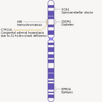
Schematic of chromosome 6, taken from
http://www.ncbi.nlm.nih.gov/books/NBK22266/#A278

THE HFE GENE

Chromosomes are depicted as shown in the image on the right. The blue bands represent clusters of genes. Chromosome six is composed of 170 million base pairs and contains about 2,000 genes. The HFE gene is just one of these genes, and is 8,000 base pairs long. There are 20 different locations along this 8,000 base pair region which will result in hemochromotosis. However, there are two locations in particular where mutations occur quite frequently, and they result in two changes in the HFE protein. These mutations are called C282Y, and H68D. 

ADDITIONAL MUTATIONS
There are at least 52 additional point mutations across the HAMP, HFE2, SLC40A1, and TFR2 genes that can all result in hemochromtosis. 

MEDICAL RELEVANCE
I mentioned in the beginning that I have hemochromotosis. However, this may not be entirely accurate. My situation is that I was screened for HH on the basis of a blood test which showed elevated iron levels. However, the genetic test showed an unusual result. Most people with HH are homozygous in that they have two copies of the defective HFE gene. My results came back as being heterozygous, with one copy of my HFE gene having both the C282Y, and H68D mutation, which my doctor says should not result in any symptoms. However, I do have at least some of the symptoms, and I would like to rule out HH as one of the causing factors. Until I started researching this disease I was unaware there were so many genes and point mutations that could result in its development. Personally, I find it interesting, but I would also like to know that my doctors have considered the possibility that I also carry a defective copy of one of the other HH genes. However, I think I am correct in saying that the probability of me having another defective copy of a mutated gene which relates to HH, but is not the HFE gene are reasonably slim due to something called conjunction fallacy, see here.

CONCLUSION
So, you now understand the basics of hemochromotosis. There will undoubtedly be much more for me to discover about this disease, and as mentioned, I plan to provide additional posts that go into details on the biochemistry of the disease. Each of these defective genes make a unique protein that misbehaves in a unique way which will very exciting to learn more about. I appreciate this is a very technical blog post, so if there were things you did not understand it's because I have not explained them well. If this is the case, feel free to leave a comment below, and I will happily review and re-write to make it better.

USEFUL MATERIAL
Original research paper Hemochromatosis types 1, 2, and 3